|
How Does the Heart Work?
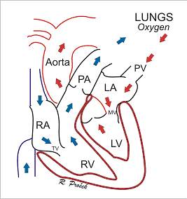
The heart is the organ responsible for pumping blood to and from all tissues of the body. The heart is divided into right and left sides. The job of the right side is to pump oxygen-deficient blood returning from the body into the lungs to be re-oxygenated, and to get rid of the carbon dioxide that the body produces. Returning from the lungs, the now-oxygenated blood enters the left side of the heart where it is pumped out into the aorta and to the body via the arterial system.
Each side of the heart also has two chambers, an upper atrium and a lower ventricle. Between the atrium and ventricle on each side lies a valve ? the tricuspid on the right and the mitral on the left ? that regulates blood flow into the chambers. As the heart pumps, these valves act as one-way gates allowing blood to flow from the atrium above to the ventricle below and preventing blood from flowing back into the atrium. From the ventricles, blood is then forced to flow out into the pulmonary artery (on the right) or the aorta (on the left) through a second series of one-way valves (the pulmonic valve and the aortic valve).
What is Degenerative Valve Disease?
Degenerative valve disease (DVD), also called endocardiosis, valvular regurgitation,
valvular insufficiency or chronic valve disease, refers to a noninfectious
degeneration of the cardiac valves. In dogs and cats, the most commonly
affected valve is the mitral valve (on the left side of the heart), followed
by the tricuspid valve (on the right side of the heart). The pulmonic
and aortic valves are rarely affected by this condition. For reasons
we don't completely understand, the mitral or tricuspid valve leaflets
can become abnormally thickened and develop a nodular appearance in some
breeds (see below). These, and other changes to the valves, impede
their ability to form a tight seal between the atrium and ventricle during
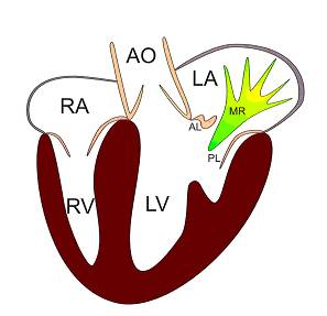
systole (contraction of the heart muscle) ? they begin to leak. As
a result, some of the blood in the ventricle now flows back into the atrium
through the leaky valve (known as regurgitation) instead of moving forward
from the ventricle into the aorta (on the left side) (Figure 2) or pulmonary
artery (on the right side) with each beat. The consequences of this
are discussed below.
What Animals are Affected by DVD?
Degenerative valve disease accounts for about 75% of cardiovascular disease in dogs. Approximately 60% of affected dogs have degeneration of the mitral valve, 30% have lesions in both the tricuspid and mitral valve leaflets, and 10% have only tricuspid valve disease. In dogs, the disease is age and breed-related, with older, small-breed dogs demonstrating a higher incidence. There is also a slight predisposition among male dogs.
Miniature poodles, Cocker Spaniels, Miniature Schnauzers, and Dachshunds
are the most commonly affected breeds. Cavalier King Charles Spaniels, another commonly affected breed, tend to develop DVD earlier in life with
a faster progression than the other small breed dogs. Terrier breeds
are also commonly seen with DVD.
Larger breeds are also affected by this disease (most commonly the mitral valve leaflets) although much less often.
Degenerative valve disease is uncommon in cats.
How Does DVD Affect your Dog?
The outcome of DVD in dogs depends on the severity of the condition. The clinical signs depend on which valve is affected.
The regurgitation of blood due to DVD causes a murmur (abnormal heart sound)
when your veterinarian listens to your dogs heart. Murmurs due to
mitral and/or tricuspid regurgitation are much more common than clinical
signs related to the disease. Many patients develop only mild and
slowly progressive valvular lesions that have no effect on the patients
quality of life.
However, when mitral regurgitation is substantial, the flow of blood back into the left atrium results in a blood volume overload inside the left atrium and left ventricle, causing these chambers to get bigger as they attempt to accommodate the extra blood. If the regurgitation is severe, then the chamber enlargement reaches a limit and the pressure inside these chambers begins to increase. If the left atrial pressures become significantly high as a result of increased blood volume, fluid within the lung vessels (which are connected to the left atrium) begins to leak out, resulting in clinical signs of congestive heart failure (also known as pulmonary edema).
Congestive heart failure due to DVD of the mitral valve usually presents as coughing, shortness of breath and rapid breathing. The generalized decrease in effective forward circulation of blood to the tissues of the body may also manifest as lethargy, exercise intolerance, lack of appetite, or weight loss.
Tricuspid valve DVD has a similar course of events as mitral valve DVD, but instead of fluid building up in the lungs (pulmonary edema), fluid builds up in body cavities ? the abdomen, the pleural space (chest cavity). Dogs with right-sided congestive heart failure tend to develop a grossly distended abdomen. This can cause some discomfort, especially when lying down, and can cause a shortness of breath, especially when sleeping or resting.
How is the Valve Disease Treated?
Treatment of DVD centers on eliminating signs of congestive heart failure.
Drugs commonly used are diuretics (furosemide, hydrochlorothiazide, spironolactone), angiotensin converting enzyme inhibitors (enalapril, benazepril, ramipril), afterload reducers (amlodipine, hydralazine), digoxin and newer drugs, such as pimobendan. With right-sided
congestive heart failure (fluid in the abdomen), repeated physical removal by your veterinarian is often the best option.
Can I Slow Down or Reverse the Progression of DVD in My Pet?
Several trials have looked at preventing DVD from progressing to the point of congestive heart failure. Unfortunately, no drugs that have been looked at have proven effective in either preventing or slowing down progression of DVD. As new drugs are developed, these will also undergo testing, so there may be drugs in the future that could help with DVD progression.
Can Diet Help?
Some animal diet manufacturers have developed heart-specific diets that are moderately restricted in salt. However, while these diets are unlikely to be harmful, they have not been shown to affect progression of the disease or control of clinical signs.
Is There Surgery to Correct DVD?
In human medicine, valve replacement or repair is a common surgical procedure. Unfortunately, surgical exposure of the mitral or tricuspid valves requires cardiopulmonary bypass. Although currently being performed by several veterinary surgeons throughout the country, bypass surgery is difficult to perform in small animals. Costs for valve repair/replacement are approximately $10,000. There are limitations on the types of patients that are suitable for surgery. If you wish to investigate the possibility of surgical correction, you should discuss this with your veterinarian. Alternative corrective procedures are in trials and may one day be available to canine patients with DVD.
Heart transplants are not an option in dogs because it would require killing a healthy dog to obtain its heart ? something that is considered unethical by the veterinary profession.
What Should You Monitor at Home?
It is important that you monitor you pets overall attitude and any change
in behavior. It may also be helpful for you to keep record of your
pets respiratory rate (number of breaths per minute) so that you will notice
increases or changes from normal breathing. Normal dogs, and dogs with well controlled heart disease usually have a
breathing rate of 1 breath every 2 to 3 seconds (20-30 breaths/min).
If breathing rates are much higher than this, or if you notice any of the
following signs, please contact a veterinarian immediately:
�E heavy, labored, or rapid breathing
�E increased coughing
�E fainting spells
�E restlessness
�E anorexia
Figure 1. Blue arrows represent deoxygenated blood flowing through the right side of the heart to the lungs, where red arrows represent oxygenated blood leaving the lungs through the left side of the heart. RA = right atrium, RV = right ventricle, TV = tricuspid valve, PA = pulmonary artery, PV = pulmonary vein, LA = left atrium, MV = mitral valve, LV = left ventricle.
Figure 2. Graphic representation of mitral valve insufficiency (regurgitation). RA = right atrium, RV = right ventricle, AO = aorta, LA = left atrium, LV = left ventricle, MR = mitral regurgitation, AL = anterior mitral leaflet, PL = posterior mitral leaflet
*Quotation from VIN |