Canine
Cystoisospora canis
Cystoisospora ohioensis
Cystoisospora neorivolta
Cystoisospora burrowsi
Feline
Cystoisospora felis
Cystoisospora rivolta
*Canine and feline Cystoisospora spp. are sometimes referred to as Isospora.
-
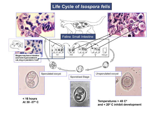
Overview of Life Cycle
- Nonsporulated (noninfective) oocysts in feces
- Sporulated (infective) oocysts in the environment
- Schizonts (asexual stages) in the small and/or large intestine
- Gametes (sexual stages) in the small and/or large intestine
- Zoites, which may be sporozoites ormerozoites, are found in extraintestinal tissues (i.e., mesenteric lymph nodes, liver, or spleen) of definitive host as well as in paratenic (transport hosts) such as mice, rats, hamsters, and other vertebrates.
-
Disease
- Coccidiosis causes diarrhea with weight loss, dehydration, and (rarely) hemorrhage
- Severely affected animals may present with anorexia, vomiting, and depression. Death is a potential outcome.
- Dogs and cats may shed oocysts in feces but remain asymptomatic.
- Intercurrent disease(s), infectious or iatrogenic immunosuppression, or the stresses of environmental changes (i.e., shipment to pet stores or relocation to pet owners) may exacerbate coccidiosis.
-
Prevalence
- Coccidial infections are common in dogs and cats.
- Published surveys indicate that coccidia are present in from 3% to 38% of dogs and 3% to 36% of cats in North America.
- Young animals are more likely than older animals to become infected with coccidia.
-
Host Associations and Transmission Between Hosts
- Canine and feline coccidia are acquired by ingestion of sporulated oocysts from contaminated environments.
- Coccidiosis is also transmitted to dogs and cats by ingestion of transport hosts (predation) containing extraintestinal stages.
- Cystoisospora spp. are rigidly host-specific. Canine coccidia will not infect felines leading to passage of oocysts in feces. The same is true for feline coccidia.
- Canine and feline Cystoisospora spp. are not known to be of zoonotic significance.
-
Prepatent Period and Environmental Factors
- Development of oocysts to infective sporulated oocysts (sporulation) does not occur above 40° C or below 20° C.
- Sporulation occurs rapidly (<16 hours) at temperatures between 30° C and 37° C.
- Sporulated oocysts are resistant to adverse environmental conditions and can survive as long as one year in moist, protected environments if they are not exposed to freezing or extremely high temperatures.
-
Site of Infection and Pathogenesis
- Developmental stages reside in either cells lining the intestinal villus (enterocytes) or cells within the lamina propria of the villus.
- Maturation and emergence of asexual and sexual stages from infected cells causes cell lysis. This damage can be especially severe when caused by species that develop within cells in the lamina propria.
- Zoites also are found in extraintestinal tissues (i.e., mesenteric lymph nodes, liver, or spleen) of definitive or paratenic hosts. These resting or latent stages are not thought to cause clinical disease.
-
Diagnosis
- Diagnosis of canine and feline coccidiosis is based on signalment, history, and clinical signs, and the structure of oocysts present in feces
- Fecal examination should be performed using centrifugal flotation and
an adequate amount of feces.
- Several genera of coccidia-like organisms may be present in canine and feline feces. It is important to differentiate them on the basis of size, state of sporulation, and presence/absence of oocysts or sporocysts.
- The presence of oocysts in feces is not, in itself, adequate proof that coccidiosis is the cause of accompanying clinical signs.
- Oocysts of Eimeria spp. are sometimes observed in canine and feline fecal samples. Dogs and cats are not hosts to Eimeria spp.; therefore these oocysts are referred to as pseudoparasites. These oocysts never reach the two-celled stage typical of Cystoisospora spp. A few two-celled Cystoisospora oocysts are often observed, even in fresh fecal samples. Additionally, oocysts of many Eimeria spp. often have oocyst wall ornamentations called micropyles or micropyle caps.
-
Treatment
- Sulfadimethoxine is the only drug that is label approved for treatment of enteritis associated with coccidiosis.
- Numerous additional drugs and drug combinations have been used with some success. (See below)
- Among the newer drugs, ponazuril appears to be effective, according to published research and user testimonials.
Treatment of Coccidiosis of Dogs and Cats
| Sulfadimethoxine |
50-60 mg/kg daily for 5-20 days (D.C) |
| Sulfaguanidine |
150 or 200 mg/kg daily for 6 days (D,C); 100-200 mg/kg every 8 hours for 5 days (D,C) |
| Furazolidone |
8-20 mg/kg once or twice daily for 5 days (D,C) |
| Trimethoprim/Sulfonamide |
Dose/length depends of sulfa; 30-60 mg/kg trimethoprim daily for 6 days in animals ≥ 4 kg; or 15-30 mg/kg trimethoprim for 6 days in animals ≤ 4 kg |
| Sulfadimethoxine/Ormetoprim |
55 mg/kg of sulfadimethoxine and 11 mg/kg of ormetaprim for 7-23 days (D) |
| Quinacrine |
10 mg/kg daily for 5 days (C) |
| Amprolium |
300 to 400 mg (total) for 5 days (D); 110-200 mg (total) daily for 7-12 days (D); 60-100 mg/kg (total) daily for 7 days (C); 1.5 tbsp (23 cc)/gal (sole water source) not to exceed 10 days (D) |
| Amprolium/Sulfadimethoxine |
150 mg/kg of amprolium and 25 mg/kg of sulfadimethoxine for 14 days (D) |
| Toltrazuril |
10-30 mg/kg daily for 1-3 days (D) |
| Diclazuril |
25 mg/kg daily for 1 day (C) |
| Ponazuril |
20 mg/kg daily for 1-3 days (D,C) |
|
*Quoation from CAPC
|

